This is observed mainly in persistent liver disease B and C, acute hepatitis brought on by hepatotropic viruses, non-hepatotropic viruses as well as drug poisoning. This video clip, captured using esophagoscopy, shows band ligation of esophageal varices. Video clip courtesy of Dan C Cohen, MD, as well as Dawn Sears, MD, Division of Gastroenterology, Scott & White Medical Care. Alcohol intake ought to highly be discouraged, specifically in people with alcoholic cirrhosis.
Liver disease accounts for about 2 million fatalities each year, making up an essential scientific trouble worldwide. Of those, 1.03 million deaths are due to advanced persistent liver disease and also its difficulties. The misnomer “portal set of three” typically has consisted of only the initial 3 frameworks, as well as was called before lymphatic vessels were discovered in the structure. It can refer both to the biggest branch of each of these vessels running inside the hepatoduodenal tendon, as well as to the smaller branches of these vessels inside the liver. With an ordinary lifetime of 5 months, hepatocytes present a remarkable capacity to regenerate complying with partial hepatectomy and also upon liver injury as a result of toxic substances, trauma, hair transplant, as well as ischemia/reperfusion.
Crosstalk In Between Hepatic Macrophages And Also Hepatocytes
Intracellular pigments are most frequent in the cytoplasm of hepatocytes. They are fine or crude granular aggregates that acquire different colorations which may need unique histochemistry research studies to establish the etiology. Number 5 reveals an algorithm for normal lobular architecture and portal development needing search for lobular injury or steatosis. Granulomas can be found in the portal region, in the parenchyma, or in both. The frequency of granulomas ranges from 2.4% to 15%, of which greater than 60% is second to systemic processes, one 3rd to main hepatic problems while 6% to 10% are idiopathic.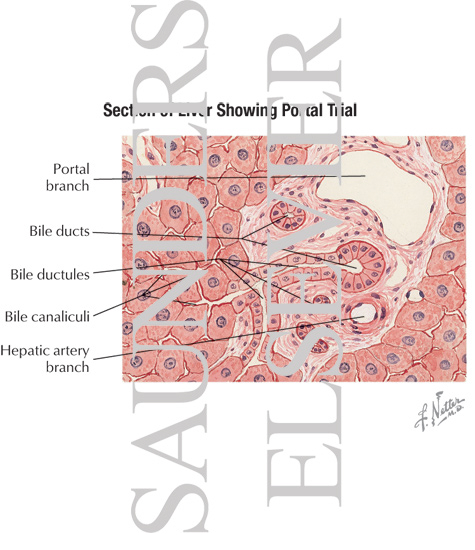
In cirrhosis, the sinusoidal links in between the portal venous system and the draining main incurable hepatic venules as well as hepatic veins are blocked or ruined. Zone 1 region contains hepatocytes that are closest to the passing through venule near the portal sets of three. Area 2 region is midway between the passing through venules as well as the incurable hepatic vein.
Framework As Well As Feature
The digestive tract likewise contains ornithine aminotransferase, and also can manufacturing ornithine from glutamate. The kidney uses glutamine as the resource of ammonium ions as a means of excretion of excess acid right into the tubular fluid. This response is a major resource of arginine synthesis as well as clarifies why arginine is not a vital amino acid in normal human beings.
- An alternate organization of hepatocytes places the line in between 2 triads at the center of a rhomboid with the ends being central veins.
- Blood goes into the lobule from the portal triad, which includes a portal blood vessel, bile air duct, hepatic artery, and also lymphatic vessels.
- In the absence of efficient urea cycle activity, just the glutamine-producing responses would certainly be energetic in the liver, hence the extreme hyperglutaminemic characterstic of urea cycle conditions.
- A raised pressure difference between systemic as well as portal blood circulation straight adds to the growth of varices.
- In siderosis because of transfusions or diet, iron down payments lie in the Kupffer cells.
Hepatocytes contain rounded cores with spread chromatin and also popular nucleoli; polyploidy is a typical attribute of these cells, with 30– 40% of the grown-up human hepatocyte showing this characteristic. Notice that a slim room is present between the endothelial cells lining the sinusoids and also the parenchymal cells. This is the area of Disse, as well as it is in continuity with the lumen of the sinusoids via small spaces between the endothelial cells that create the wall surface of the sinusoids. The connective tissue septae invaginating from the capsule mark hepatic lobules, the structural unit of the liver.
What Makes The Portal Blood Vessel?
Bile produced by hepatocytes contains water, bile salts, mucin and pigments, cholesterol, esterified and nonesterified fatty acids, and also inorganic salts and has a pH of 7.1– 7.3. Bile is vital for fat emulsification in the intestine that assists in the absorption of lipids as well as lipid-soluble vitamins. It is also a tank of alkali and plays a vital function in neutralization of acid chime from the tummy. Most of the secreted bile salts are reabsorbed in the intestine and used once more for secretion. In people, hepatocytes synthesize about 200– 500mg of bile acids daily to change for their day-to-day loss in the feces.
Nonalcoholic steatohepatitis is ending up being a significant source of liver cirrhosis in the United States as liver disease C is becoming a significant cause of liver cirrhosis worldwide. Enhanced portal pressure adds to increased varix size as well as decreased varix wall thickness, therefore bring about enhanced variceal wall surface tension. Rupture happens when the wall stress exceeds the elastic restrictions of the variceal wall surface.
Delayed venous phase of a careful typical hepatic angiogram shows the portal vein, with dental filling of the left gastric capillary brought on by retrograde flow feeding gastric as well as lower esophageal varices. The final diagnosis was liver disease C cirrhosis, hepatocellular cancer of the left hepatic lobe, as well as portoarterial fistula. In general, alcoholic liver illness as well as viral hepatitis are one of the most usual causes for esophageal varices in both sexes. In men with esophageal varices, alcoholic liver illness and viral hepatitis are normally the reason. This is based upon the monitoring that glucagon-null computer mice display a modified gene expression pattern, especially in the periportal area. Glutamine synthase is a classical marker for the pericentral area, and also its expression changes from remaining in the inner three cell layers in wild-type mice to extending out almost two times as much in glucagon-null mice.